Radiation Sources:
Two of the most commonly used sources of radiation in industrial radiography are x-ray generators and gamma ray sources. Industrial radiography is often subdivided into "X-ray Radiography" or "Gamma Radiography", depending on the source of radiation used.
Gamma Radiography:
Gamma rays are similar to X- Rays except that they have much shorter wavelength and differ in their origin. Gamma rays are emitted from the nucleus itself during the process of radioactivity. Gamma rays are produced by a radioisotope. A radioisotope has unstable nuclei that do not have enough binding energy to hold the nucleus together. The spontaneous breakdown of an atomic nucleus resulting in the release of energy and matter is known as radioactive decay.
Most of the radioactive material used in industrial radiography is artificially produced. This is done by subjecting stable material to a source of neutrons in a special nuclear reactor. This process is called activation. Two most commonly used gamma ray sources in industrial radiography are iridium 192 and cobalt 60.
X-Ray RADIOGRAPHY
X - Rays are produced whenever high energy electrons suddenly give up energy. This can be done either by accelerating electrons to a high speed and then stopping them suddenly, or by these high speed electrons striking others and knocking them out of their normal positions. When these dislodged electrons fall back into place they give off X-Rays. X-rays are produced by establishing a very high voltage between two electrodes, called the anode and cathode. To prevent arcing, the anode and cathode are located inside a vacuum tube, which is protected by a metal housing. The cathode contains a small filament much the same as in a light bulb. Current is passed through the filament which heats it. The heat causes electrons to be stripped off. The high voltage causes these "free" electrons to be pulled toward a target material (usually made of tungsten) located in the anode. The electrons impact against the target. This impact causes an energy exchange which causes x-rays to be created.
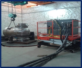
COBALT - 60 RADIOGRAPHY
Cobalt-60 is a preferred source for the radiography of steel thickness of about 75mm to 200 mm. We have world's latest and sophisticated gamma ray projectors and proud to say that we are the only private agency in India having a maximum number of Cobalt -60 (TECH OPS) exposure devices.
IRIDIUM-192 RADIOGRAPHY
Iridium 192 Radiography: Iridium -192 is used for radiography of steel thickness of about 6mm to 75 mm. We have Exposure devices (SPEC2T & TECH OPS) that are all imported and the latest.
BETATRON RADIOGRAPHY
Betatron is basically a combination of an electromagnet and a transformer designed to guide and accelerate electrons in a circular orbit to very high energies. The toroidal type of hot cathode high vaccum X-ray tube commonly used in betatron is capable of injecting and energizing electrons to many millions of volts before striking the target to produce X ray. Betatron of this type have been constructed to generate X-rays at energies ranging from 15 to 100MeV.The average beam current is on the order of 1 to 3µA.The Focal spot of the target is usually less than 1mm(0.04in) in diameter. Commercially available betatrons are capable of radiographing steel in the range of 5to 41 mm.